Your Cart is Empty


 |
This simple 10-minute course into melanoma diagnosis using the 7-point checklist will give you an overview of pattern analysis. The 7-point checklist provides a simplification of standard pattern analysis because of the low number of features, or criteria, to identify. As with the ABCD rule, it can be easily learned and applied and has proven to be reliable in diagnosing melanoma. |
| Major ELM Criteria | Score |
|---|---|
| Atypical pigment network | 2 |
| Gray-blue areas | 2 |
| Atypical vascular pattern | 2 |
| Minor ELM Criteria | Score |
| Radial streaming (streaks) | 1 |
| Irregular diffuse pigmentation (blotches) | 1 |
| Irregular dots and globules | 1 |
| Regression pattern | 1 |
The score for criterion presence is determined on the basis of the odds ratio.
Odds ratios measure the capacity of each criterion of increasing the probability of a positive melanoma diagnosis.
By simple addition of the criteria scores, a minimum total score of 3 is required for the diagnosis of melanoma.
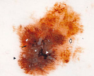
Definition
Prominent (hyperpigmented or broad) and irregular network
Histological Correlates
Hyperpigmented or broadened rete ridges with irregular shape or distribution
Sample Lesion
Irregular and prominent (atypical) pigment network (white arrow)
(7-point score: 2)
streaks (black arrowhead)
(score: 1)
blotches (white arrowhead)
(score: 1)
irregular dots and globules (black arrow)
(score: 1)
Cutaneous melanoma, 0.45 mm thick
(original magnification 10x)
TOTAL SCORE: 5

Definition
Prominent (hyperpigmented or broad) and irregular network
Histological Correlates
Hyperpigmented or broadened rete ridges with irregular shape or distribution
Sample Lesion
Irregular, but discrete, pigment network (score: 0)
Regression pattern (peppering within depigmented areas, asterisk)
The lesion is asymmetrical , with 4 colors and 3 dermoscopic structures
irregular dots and globules (black arrow)
(score: 1)
Atypical Nevus
(original magnification 10x)
TOTAL SCORE: 1

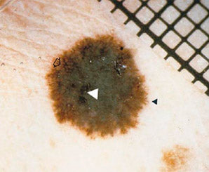
Definition
Irregular, confluent, gray-blue to whitish blue diffuse pigmentation not associated with red-blue lacunes or maple leaf pigmentation
Histological Correlates
Pigmented melanophages or melanocytes of midreticular dermis location
Sample Lesion
with a prevalence of gray-blue areas (curved black arrow) (score: 2)
An atypical pigment network (white arrow
streaks (black arrowhead)
blotches (white arrowhead)
irregular dots and globules (black arrow)
Cutaneous melanoma, 1.0 mm thick
(original magnification 10x)
TOTAL SCORE: 7

Definition
Linear, dotted, or globular red structures irregularly distributed outside areas of regression and associated with other melanocytic pigment patterns
Histological Correlates
Neovascularization or vascularized nests of amelanotic cells
Sample Lesion
Atypical (dotted and globular) vascular pattern (long black arrowheads) (score: 2)
Gray-blue areas (curved black arrow)
treaks (short black arrowhead)
Cutaneous melanoma, 0.8 mm thick
(original magnification 10x)
TOTAL SCORE: 5

Definition
Linear, dotted, or globular red structures irregularly distributed outside areas of regression and associated with other melanocytic pigment patterns
Histological Correlates
Neovascularization or vascularized nests of amelanotic cells
Sample Lesion
Commalike vessels, commonly associated with dermal papillae in compound and dermal nevi(black arrowhead) (score: 0)
Compound melanocytic nevus
(original magnification 10x)
TOTAL SCORE: 5

Definition
Radially and asymmetrically arranged linear or bulbous extensions at the edge of the lesion
Histological Correlates
Confluent radial junctional nests of melanocytes
Sample Lesion
Streaks (black arrowhead)
Irregular dots and globules (thin black arrow)
Regression pattern consists of white areas (asterisk) and peppering (thick black arrow) (score: 1)
Cutaneous melanoma, 0.6 mm thick
(original magnification 10x)
TOTAL SCORE: 3
Definition
Brown, gray, and black areas of diffuse pigmentation with irregular shape or distribution and abrupt end
Histological Correlates
Hyperpigmentation throughout all levels of the epidermis or upper dermis (in melanocytes or melanophages)
Sample Lesion
Blotches (white arrowhead)
Presence of atypical pigment network (white arrow)
Relatively symmetrical lesion with abrupt cutoff of pigment pattern, 3 colors, and 4 structures
streaks (black arrowhead)
Cutaneous melanoma, 0.3 mm thick
(original magnification 10x)
TOTAL SCORE: 4

Definition
Black, brown, or blue round structures irregularly distributed within the lesion
Histological Correlates
Aggregates of pigment of stratum corneum, junctional, or dermis location
Sample Lesion
Irregular dots and globules (black arrows)
Blotches (white arrowhead)
Streaks (black arrowhead
Regression pattern (white areas, thick black arrows)
Two lesions with similar silhouettes and distribution of colors and structures (see next page)
Compound melanocytic nevus
(original magnification 10x)
TOTAL SCORE: 2

Definition
Black, brown, or blue round structures irregularly distributed within the lesion
Histological Correlates
Aggregates of pigment of stratum corneum, junctional, or dermis location
Sample Lesion
Irregular dots and globules (black arrows)
Blotches (white arrowhead)
Streaks (black arrowhead
Regression pattern (white areas, thick black arrows)
Two lesions with similar silhouettes and distribution of colors and structures (see previous page)
Cutaneous melanoma, 0.45 mm thick
(original magnification 10x)
TOTAL SCORE: 2

Definition
White scarlike depigmentation or "peppering" (speckled multiple blue-gray dots within a hypodepigmented area)irregularly distributed within the lesion
Histological Correlates
Areas of loss of pigmentation and fibroplasia, with scattered dermal melanophages
Sample Lesion
Regression pattern (black arrows)
Irregular, discrete pigment network
The lesion is asymmetrical, with abrupt cutoff of pigment pattern, 4 colors, and 3 dermoscopic structures
Atypical melanocytic nevus
(original magnification 10x)
TOTAL SCORE: 1
Stay on top of the latest developments in dermoscopy and beyond